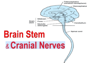
Brain Stem & Cranial Nerves 1
Cornell notes
✒️ Title: Brainstem Anatomy and Cranial Nerves
🌟 Cues
- Brainstem regions: Midbrain, Pons, Medulla
- Cranial nerves associated with brainstem
- Functions of brainstem structures
- Relation of cranial nerves to sensory and motor functions
🗒️ Notes
Brainstem Overview: The brainstem consists of the midbrain, pons, and medulla. Each section is crucial for relaying signals between the brain and the spinal cord, and for controlling basic life functions.
Midbrain: Located between the diencephalon and pons, the midbrain contains the cerebral aqueduct, which divides it into the cerebral peduncles and the tectum. The superior colliculi handle visual reflexes, and the inferior colliculi are part of the auditory pathway.
Pons: The pons is a bridge between the midbrain and medulla, housing the trigeminal, facial, vestibulocochlear, and abducens nerves. Its basilar part contains important structures like the middle cerebellar peduncle and facial colliculus.
Medulla Oblongata: The medulla transitions into the spinal cord at the foramen magnum. It controls essential functions like heart rate and respiration. Notable structures include the pyramids, inferior olivary nucleus, and rootlets of several cranial nerves (IX, X, XI).
Cranial Nerves: There are 12 cranial nerves, categorized as sensory, motor, or both. Specific nerves such as the oculomotor (III), trochlear (IV), and abducens (VI) control eye movements, while the facial (VII) and trigeminal (V) are responsible for facial sensations and expressions.
📝 Summary
The brainstem plays a critical role in integrating and relaying information between the brain and body. Each section—the midbrain, pons, and medulla—has specialized structures essential for sensory processing, motor control, and maintaining vital functions. Additionally, the cranial nerves originating from the brainstem perform vital sensory and motor tasks, such as eye movement, facial expressions, and control of visceral organs.

